LIVER DISEASE
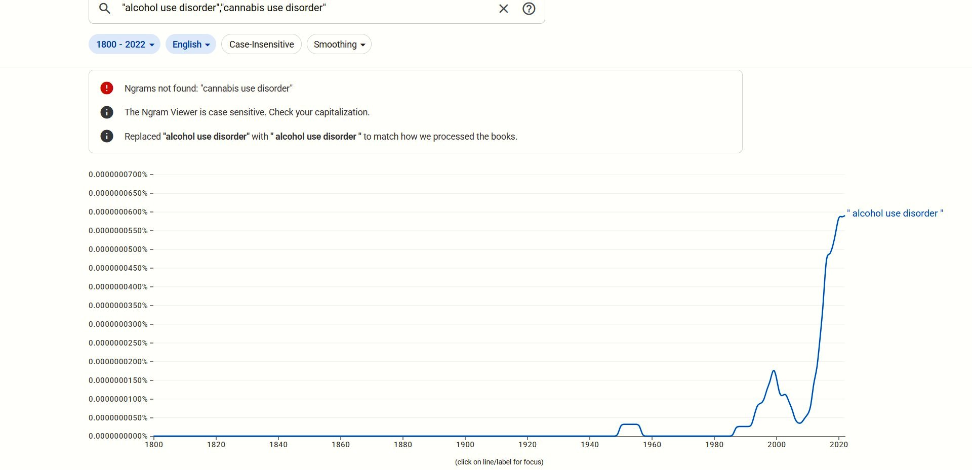
Following the runaway success of "alcohol use disorder" attempts to
set up a rival disorder - "cannabis use disorder" - met with little
success:
Sticking to the script, Fakhoury et al (2025) found that alcoholics
are less likely to die from alcoholic liver disease (ALD) by abusing
cannabis:
"Using the TriNetX US Collaborative Network, we identified adult
patients with AUD between 2010 and 2022. Three cohorts were
constructed: cannabis use disorder (CUD), cannabis users without
cannabis abuse or dependence (CU) and non-cannabis users (non-CU).
Outcomes included ALD, hepatic decompensation and composite all-cause
mortality over 3 years. Incidence and hazard ratios were calculated
using Kaplan-Meier analysis and Cox regression.
"Results: After matching, 33 114 patients were included in each of the
CUD and non-CU groups. Compared to non-CU, CUD was associated with a
lower risk of ALD (HR 0.60, 95% CI 0.53-0.67; p < 0.001), hepatic
decompensation (HR 0.83, 95% CI 0.73-0.95; p =0.005) and all-cause
mortality (HR 0.86, 95% CI 0.80-0.94; p < 0.001) among individuals
with AUD. Although CU was associated with lower risks of ALD, its
risks of hepatic decompensation and all-cause mortality were similar
to those of the non-CU cohort with AUD."
Notice how in these unpleasant
results "similar" means the same as 40%, 17%, and 14% lower risks.
https://pubmed.ncbi.nlm.nih.gov/41117396/ [5505]
Not all liver disease is exclusively
alcohol-related. Sometimes it's connected with antipsychotic drugs.
However such mechanisms are not exclusive to those administered
psychiatric drugs. How would your liver know if you are psychotic or
not? According to Váquez-Bourgon et al (2019) at Universidad de
Cantabria, Santander, Spain:
"Increased incidence of obesity and excess weight lead to an increased
incidence of non-alcoholic fatty liver disease (NAFLD). Recent
evidence indicates a protective effect of cannabis consumption on
weight gain and related metabolic alterations in psychosis patients.
Overall, patients are at greater risk of presenting fatty diseases,
such as NAFLD, partly due to lipid and glycemic metabolic
disturbances. However, there are no previous studies on the likely
effect of cannabis on liver steatosis. We aimed to explore if cannabis
consumption had an effect on hepatic steatosis, in a sample of
first-episode (FEP) non-affective psychosis.
"Material and methods
A total of 390 patients were evaluated at baseline and after 3 years
of initiating the antipsychotic treatment. Anthropometric measurements
and liver, lipid, and glycemic parameters were obtained at both time
points. All but 6.7% of patients were drug-naïve at entry, and
they self-reported their cannabis use at both time points. Liver
steatosis and fibrosis were evaluated through validated clinical
scores (Fatty Liver Index [FLI], Fibrosis-4 [FIB-4], and NAFLD).
"Results
At 3-year follow-up, cannabis users presented significantly lower FLI
scores than non-users (F = 13.874; p < .001). Moreover, cannabis
users less frequently met the criteria for liver steatosis than
non-users (X2 = 7.97, p = .019). Longitudinally, patients maintaining
cannabis consumption after 3 years presented the smallest increment in
FLI over time, which was significantly smaller than the increment in
FLI presented by discontinuers (p = .022) and never-users (p = .016).
No differences were seen in fibrosis scores associated with cannabis.
"Conclusions
Cannabis consumption may produce a protective effect against liver
steatosis in psychosis, probably through the modulation of
antipsychotic-induced weight gain."
https://www.sciencedirect.com/science/article/abs/pii/S0278584619301393
[541]
Castro and Abdel Bermdez-del Sol (2025) present an alternative name
for NAFLD:
"Potential associations have been investigated between metabolic
dysfunction-associated steatotic liver disease (MASLD), formerly known
as non-alcoholic fatty liver disease, and cannabis use. This study
aimed to determine the association between cannabis use frequency and
MASLD. Up to January 2025, the evidence from PubMed, Scopus, and Web
of Science was synthesized in this systematic review and
meta-analysis, which was registered in PROSPERO (CRD42025025065) and
followed the Preferred Reporting Items for Systematic reviews and
Meta-Analyses (PRISMA) guidelines. Of the 711 initial records, 11
observational studies involving 5,968,702 individuals met the
inclusion criteria. A pooled analysis revealed that cannabis use was
associated with a reduced risk of hepatic steatosis (OR = 0.58; 95%
CI: 0.42-0.81; p = 0.002; I = 97%). The subgroup analysis revealed a
protective association for past users (OR = 0.84; 95% CI: 0.77-0.93)
and occasional users (OR = 0.35; 95% CI: 0.20-0.64), with no
significant association observed for frequent users. The study
revealed that cannabis users exhibited a decline in both the fatty
liver index (mean difference (MD) = -11.02) and the BMI (MD = -1.89
kg/m). However, the findings did not show any statistically
significant changes in liver fat (%), transaminases (aspartate
aminotransferase and alanine aminotransferase), and triglycerides. A
risk-of-bias assessment identified notable methodological limitations.
Overall, the findings suggest a strong association between cannabis
use and MASLD, though causality cannot be established."
https://www.researchgate.net/profile/Nestor-Quinapanta-Castro/publication/395836766_A_Systematic_Review_and_Meta-Analysis_of_Cannabis_Use_Frequency_and_Metabolic_Dysfunction-Associated_Steatotic_Liver_Disease_Scapegoat_or_Healer/links/68d5556cf3032e2b4be32fd8/A-Systematic-Review-and-Meta-Analysis-of-Cannabis-Use-Frequency-and-Metabolic-Dysfunction-Associated-Steatotic-Liver-Disease-Scapegoat-or-Healer.pdf?_tp=eyJjb250ZXh0Ijp7ImZpcnN0UGFnZSI6InB1YmxpY2F0aW9uIiwicGFnZSI6InB1YmxpY2F0aW9uIn19
[5537]
According to the BMJ:
"Nonalcoholic hepatic
steatosis, or nonalcoholic fatty liver disease (NAFLD), is one of the
most common causes of chronic liver disease in the developed world.
"It is a spectrum of disease,
ranging from hepatic fat accumulation without inflammation to
steatohepatitis, fibrosis, cirrhosis, and end-stage liver disease.
"NAFLD is considered the
hepatic manifestation of metabolic syndrome and is associated with
obesity, dyslipidemia, and type 2 diabetes mellitus.
"The diagnosis of nonalcoholic
fatty liver disease is based on exclusion of other etiologies of
hepatic steatosis, such as alcohol use, and supportive laboratory
tests and imaging. Liver biopsy and histology is the gold standard for
diagnosis, and is performed for patients at higher risk of fibrosis or
steatohepatitis."
and
"Hepatic steatosis occurs when
intrahepatic fat is ≥5% of liver weight.
"Nonalcoholic fatty liver
disease (NAFLD) is evidence of hepatic steatosis (imaging or
histologic) in the absence of secondary causes of hepatic fat
accumulation, such as significant alcohol consumption.
"NAFLD can be categorized as
nonalcoholic fatty liver (NAFL) or nonalcoholic steatohepatitis
(NASH), depending on histologic features. NAFL is the presence of
hepatic steatosis without evidence of hepatocellular injury in the
form of hepatocyte ballooning. NASH is the presence of hepatic
steatosis and inflammation with hepatocyte injury (e.g., ballooning),
with or without fibrosis. This distinction is important for prognosis
because NASH may progress to cirrhosis and liver failure. The risk of
NAFL progressing to cirrhosis or liver failure is minimal.
"To more accurately reflect the
pathogenesis of fatty liver, a new nomenclature of metabolic
associated fatty liver disease (MAFLD) has been suggested."
https://bestpractice.bmj.com/topics/en-us/796 [1968]
After five years of ZPPPD-inspired
liver damage, the benefits of cannabis could be inferred from studies
of endocannabinoid action. In a groundbreaking step, Osei-Hyiaman et
al first demonstrated that CB1R deficiency in mice conveys a protective
effect against diet-induced hepatic steatosis, independent of caloric
intake, in "Endocannabinoid activation at hepatic CB1 receptors
stimulates fatty acid synthesis and contributes to diet-induced
obesity" (2005):
"Endogenous cannabinoids acting at CB(1) receptors stimulate appetite,
and CB(1) antagonists show promise in the treatment of obesity. CB(1)
(-/-) mice are resistant to diet-induced obesity even though their
caloric intake is similar to that of wild-type mice, suggesting that
endocannabinoids also regulate fat metabolism. Here, we investigated
the possible role of endocannabinoids in the regulation of hepatic
lipogenesis. Activation of CB(1) in mice increases the hepatic gene
expression of the lipogenic transcription factor SREBP-1c and its
targets acetyl-CoA carboxylase-1 and fatty acid synthase (FAS).
Treatment with a CB(1) agonist also increases de novo fatty acid
synthesis in the liver or in isolated hepatocytes, which express
CB(1). High-fat diet increases hepatic levels of the endocannabinoid
anandamide (arachidonoyl ethanolamide), CB(1) density, and basal rates
of fatty acid synthesis, and the latter is reduced by CB(1) blockade.
In the hypothalamus, where FAS inhibitors elicit anorexia, SREBP-1c
and FAS expression are similarly affected by CB(1) ligands. We
conclude that anandamide acting at hepatic CB(1) contributes to
diet-induced obesity and that the FAS pathway may be a common
molecular target for central appetitive and peripheral metabolic
regulation."
http://www.jci.org/articles/view/23057/files/pdf [3962]
Even without alcoholism, liver
disease will knock nearly three years off your life:
"In this nationwide
population-based cohort, all patients with NAFLD diagnosis and without
baseline CVD (ascertaining from the Swedish National Patient Register
from 1987 to 2016, n = 10,023) were matched 10:1 on age, sex, and
municipality to individuals from the general population (controls, n =
96,313). CVD diagnosis and mortality were derived from national
registers. Multistate models and flexible parametric survival models
were used to estimate adjusted hazard ratios (aHRs) for CVD risk and
loss in life expectancy due to NAFLD. We identified 1037 (10.3%) CVD
events in patients with NAFLD and 4041 (4.2%) in controls. CVD risk
was 2.6-fold higher in NAFLD compared with controls (aHR = 2.61, 95%
CI = 2.362.88) and was strongest for nonfatal CVD (aHR = 3.71, 95% CI
= 3.294.17). After a nonfatal CVD event, the risk for all-cause
mortality was similar between patients with NAFLD and controls (aHR =
0.89, 95% CI = 0.641.25). Life expectancy in patients with NAFLD was,
on average, 2.8 years lower than controls, with the highest loss of
life-years when NAFLD was diagnosed in middle age (4060 years).
"Conclusions
NAFLD was associated with a
higher risk of nonfatal CVD but did not affect post-CVD mortality
risk. Patients diagnosed with NAFLD have a lower life expectancy than
the general population."
As for cardiovascular events in
those with a liver diagnosis:
"Patients with NAFLD and
cirrhosis at baseline had higher rates of all CVD events in the first
model (HR 7.92, 95% CI = 4.7113.3), although this estimate was lower
after adjustments for CVD risk factors (aHR 2.56, 95% CI = 1.315.01)."
and
"Several observations can be
made from this large nationwide cohort study set in a secondary or
tertiary setting. First, we found an elevated risk of nonfatal CVD
events in patients with NAFLD compared with matched controls. Second,
patients with cirrhosis had a higher CVD risk than controls, but not
compared to patients with noncirrhotic NAFLD. Third, while NAFLD was
associated with increased overall mortality, no increased mortality
was observed in patients with NAFLD with incident CVD compared to
matched controls who had also experienced a nonfatal CVD event.
Finally, the overall loss of life expectancy in patients with NAFLD
was about 3 years, which was affected by age and clinical setting at
diagnosis. LEL [loss of expectancy in life] was highest in
hospitalized patients and when the diagnosis of NAFLD was made at
middle age, whereas no apparent loss in life expectancy was observed
for those aged ≥80."
https://aasldpubs.onlinelibrary.wiley.com/doi/10.1002/hep.32519
[1967]
In 2017's "Cannabis use is
associated with reduced prevalence of non-alcoholic fatty liver
disease: A cross-sectional study", Adejumo et al... "...conducted a
population-based case-control study of 5,950,391 patients using the
2014 Healthcare Cost and Utilization Project (HCUP), Nationwide
Inpatient Survey (NIS) discharge records of patients 18 years and
older. After identifying patients with NAFLD (1% of all patients), we
next identified three exposure groups: non-cannabis users (98.04%),
non-dependent cannabis users (1.74%), and dependent cannabis users
(0.22%). We adjusted for potential demographics and patient related
confounders and used multivariate logistic regression (SAS 9.4) to
determine the odds of developing NAFLD with respects to cannabis use.
Our findings revealed that cannabis users (dependent and nondependent)
showed significantly lower NAFLD prevalence compared to non-users
(AOR: 0.82[0.760.88]; p<0.0001). The prevalence of NAFLD was 15%
lower in non-dependent users (AOR: 0.85[0.790.92]; p<0.0001) and
52% lower in dependent users (AOR: 0.49 [0.360.65]; p<0.0001).
Among cannabis users, dependent patients had 43% significantly lower
prevalence of NAFLD compared to non-dependent patients (AOR:
0.57[0.420.77]; p<0.0001). Our observations suggest that cannabis
use is associated with lower prevalence of NAFLD in patients. These
novel findings suggest additional molecular mechanistic studies to
explore the potential role of cannabis use in NAFLD development."
https://journals.plos.org/plosone/article/file?id=10.1371/journal.pone.0176416&type=printable
[2772]
In 2020 Collaborators of the
ANRS CO13 HEPAVIH Study Group report:
"Mortality among individuals
co-infected with HIV and hepatitis C virus (HCV) is relatively high.
We evaluated the association between psychoactive substance use and
both HCV and non-HCV mortality in HIV/HCV co-infected patients in
France, using Fine and Gray's competing-risk model adjusted for
socio-demographic, clinical predictors and confounding factors, while
accounting for competing causes of death. Over a 5-year median
follow-up period, 77 deaths occurred among 1028 patients.
Regular/daily cannabis use, elevated coffee intake, and not currently
smoking were independently associated with reduced HCV-mortality
(adjusted sub-hazard ratio [95% CI] 0.28 [0.10-0.83], 0.38
[0.15-0.95], and 0.28 [0.10-0.79], respectively). Obesity and severe
thinness were associated with increased HCV-mortality (2.44
[1.00-5.93] and 7.25 [2.22-23.6] versus normal weight, respectively).
Regular binge drinking was associated with increased non-HCV-mortality
(2.19 [1.10-4.37]). Further research is needed to understand the
causal mechanisms involved. People living with HIV/HCV co-infection
should be referred for tobacco, alcohol and weight control
interventions and potential benefits of cannabis-based therapies
investigated."
https://pubmed.ncbi.nlm.nih.gov/31286317/ [3385]
From the perspective of organ
damage from alcohol, Yan et al set out to settle the vexed question of
whether it is a good idea to add cannabis to alcohol. Reporting in the
Journal of Biochemical and Molecular Toxicology:
"Cannabinoids (CBs) are
psychoactive compounds, with reported anticancer, anti-inflammatory,
and anti-neoplastic properties. The study was aimed at assessing the
hepatoprotective effects of CB against ethanol (EtOH)-induced liver
toxicity in rats. The animals were divided into seven groups: control
(Group I) and Group II were treated with 50% ethanol (EtOH
5 mg/kg). Groups III, IV, and VI were treated with (EtOH + CB
10 mg/kg), (EtOH + CB 20 mg/kg), and (EtOH + CB
30 mg/kg), respectively. Groups V and VII consisted of animals
treated with 20 and 30 mg/kg, of CB, respectively. Biochemical
analysis revealed that Group IV (EtOH + CB 20 mg/kg) had
reduced levels of ALTalanine transferase, ASTaspartate
aminotransferase, ALPalanine peroxidase, MDAmalondialdehyde and
increased levels of GSH-reduced glutathione. Histopathological
analysis of liver and kidney tissues showed that EtOH + CB (20 and
30 mg/kg) treated animal groups exhibited normal tissue
architecture similar to that of the control group. ELISA
[enzyme-linked immunosorbent assay] revealed that the inflammatory
markers were reduced in the animal groups that were treated with EtOH
+ CB 20 mg/kg, in comparison to the animals treated only with
EtOH. The mRNA expression levels of COX-2, CD-14, and MIP-2 showed a
remarkable decrease in EtOH + CB treated animal groups to control
groups. Western blot analysis revealed that CB downregulated
p38/JNK/ERK thereby exhibiting its hepatoprotective property by
inhibiting mitogen-activated protein kinase pathways. Thus, our
findings suggest that CB is a potential candidate for the treatment of
alcohol-induced hepatotoxicity.
https://onlinelibrary.wiley.com/doi/abs/10.1002/jbt.23260 [1859]
El Moneim Hussein et al of
Alexandria University in Egypt used a different experimental model of
liver damage in "Protective and therapeutic effects of cannabis plant
extract on liver cancer induced by dimethylnitrosamine in mice"
(2019), finding that the Group III mice, who received a cannabis
extract with a ratio of ~2:1 THC:CBD pre-treatment in advance of the
DMNA did better than those who received cannabis with or after the
treatment, concluding:
"...exposure to DMNA plays a
role in pathogenesis of liver disease leading to carcinogenicity and
causes disturbances in the activities of mice liver enzymes while
cannabis causes a partial improvement in these enzymes. The protective
effect of cannabis extract is more pronounced than other groups and
this is demonstrated in group III. Cannabinoids might exert their
anti-tumor effects by direct induction of apoptosis and can decrease
telomerase activity by inhibiting the expression of hTERT [telomerase
reverse transcriptase] gene. Coordination between inhibition of
telomerase activity and induction of apoptosis might be a potential
therapeutic agent for cancer treatment."
https://www.tandfonline.com/doi/full/10.1016/j.ajme.2014.02.003
[4073]
Glutathione exists in reduced
(GSH) and oxidized (GSSG) states. The ratio of reduced glutathione to
oxidized glutathione within cells is a measure of cellular oxidative
stress where increased GSSG-to-GSH ratio is indicative of greater
oxidative stress. In healthy cells and tissue, more than 90% of the
total glutathione pool is in the reduced form (GSH), with the
remainder in the disulfide form (GSSG).
In the reduced state, the thiol
group of cysteinyl residue is a source of one reducing equivalent.
Glutathione disulfide (GSSG) is thereby generated. The oxidized state
is converted to the reduced state by NADPH. This conversion is
catalyzed by glutathione reductase:
NADPH + GSSG + H2O → 2 GSH
+ NADP+ + OH−
https://en.wikipedia.org/wiki/Glutathione [1922]
In 2011 Vara et al reported the
"Anti-tumoral action of cannabinoids on hepatocellular carcinoma: role
of AMPK-dependent activation of autophagy":
"Hepatocellular carcinoma (HCC)
is the third cause of cancer-related death worldwide. When these
tumors are in advanced stages, few therapeutic options are available.
Therefore, it is essential to search for new treatments to fight this
disease. In this study, we investigated the effects of cannabinoids--a
novel family of potential anticancer agents--on the growth of HCC. We
found that Δ(9)-tetrahydrocannabinol (Δ(9)-THC, the main
active component of Cannabis sativa) and JWH-015 (a cannabinoid
receptor 2 (CB(2)) cannabinoid receptor-selective agonist) reduced the
viability of the human HCC cell lines HepG2 (human hepatocellular
liver carcinoma cell line) and HuH-7 (hepatocellular carcinoma cells),
an effect that relied on the stimulation of CB(2) receptor. We also
found that Δ(9)-THC- and JWH-015-induced autophagy relies on
tribbles homolog 3 (TRB3) upregulation, and subsequent inhibition of
the serine-threonine kinase Akt/mammalian target of rapamycin C1 axis
and adenosine monophosphate-activated kinase (AMPK) stimulation.
Pharmacological and genetic inhibition of AMPK upstream kinases
supported that calmodulin-activated kinase kinase β was
responsible for cannabinoid-induced AMPK activation and autophagy. In
vivo studies revealed that Δ(9)-THC and JWH-015 reduced the
growth of HCC subcutaneous xenografts, an effect that was not evident
when autophagy was genetically of pharmacologically inhibited in those
tumors. Moreover, cannabinoids were also able to inhibit tumor growth
and ascites in an orthotopic model of HCC xenograft. Our findings may
contribute to the design of new therapeutic strategies for the
management of HCC."
https://www.nature.com/articles/cdd201132.pdf [3691]
In 2024 Tan et al reported:
"Cannabidiol (CBD) has antioxidant and anti-inflammatory activities.
However, the anti-tumor effect of CBD on hepatocellular carcinoma
(HCC) remains unclear. Here, we investigated whether CBD displays
anti-tumorigenic effects in HCC cells and whether it could reduce
tumorigenesis and metastases in vivo. First, this study treated HCC
cells with different concentrations of CBD, followed by analyzing the
changes in the proliferative, apoptotic, migratory and invasive
abilities. The effects of CBD on the growth and metastasis of HCC
cells in vivo were verified by tumorigenesis and metastasis assays.
Subsequently, the target genes of CBD were predicted through the
SwissTarget website and the genes differentially expressed in cells
after CBD treatment were analyzed by microarray for intersection. The
enrichment of the pathways after CBD treatment was analyzed by KEGG
enrichment analysis, followed by western blot validation. Finally,
rescue assays were used to validate the functions of genes as well as
pathways in the growth and metastasis of HCC cells. A significant
weakening of the ability of HCC cells to grow and metastasize in vitro
and in vivo was observed upon CBD treatment. Mechanistically, CBD
reduced GRP55 expression in HCC cells, along with increased TP53
expression and blocked MAPK signaling activation. In CBD-treated
cells, the anti-tumor of HCC cells was restored after overexpression
of GRP55 or deletion of TP53. CBD inhibits the MAPK signaling
activation and increases the TP53 expression by downregulating GRP55
in HCC cells, thereby suppressing the growth and metastasis of HCC
cells."
https://pubmed.ncbi.nlm.nih.gov/38825256/
[6844]
Adejumo et al have performed several studies on the incidence of
hepatic disorders and cannabis:
In 2018's "Cannabis use is
associated with reduced prevalence of progressive stages of alcoholic
liver disease" the aim was to
"...determine the effects of
cannabis use on the incidence of liver disease in individuals who
abuse alcohol.
"Methods: We analysed the 2014
Healthcare Cost and Utilization Project-Nationwide Inpatient Sample
(NIS) discharge records of patients 18 years and older, who had a past
or current history of abusive alcohol use (n = 319 514). Using the
International Classification of Disease, Ninth Edition codes, we
studied the four distinct phases of progressive ALD with respect to
three cannabis exposure groups: non-cannabis users (90.39%),
non-dependent cannabis users (8.26%) and dependent cannabis users
(1.36%). We accounted for the complex survey sampling methodology and
estimated the adjusted odds ratio (AOR) for developing AS, AH, AC and
HCC with respect to cannabis use (SAS 9.4).
"Results: Our study revealed
that among alcohol users, individuals who additionally use cannabis
(dependent and non-dependent cannabis use) showed significantly lower
odds of developing AS, AH, AC and HCC [alcoholic steatosis,
steatohepatitis, fibrosis-cirrhosis, hepatocellular carcinoma] (AOR:
0.55 [0.48-0.64], 0.57 [0.53-0.61], 0.45 [0.43-0.48] and 0.62
[0.51-0.76]). Furthermore, dependent users had significantly lower
odds than non-dependent users for developing liver disease.
"Conclusions: Our findings
suggest that cannabis use is associated with a reduced incidence of
liver disease in alcoholics."
https://pubmed.ncbi.nlm.nih.gov/29341392/ [2771]
In another 2018 paper Adejumo
et al, starting with 188,333 records, also looked at the effect of
cannabis use on chronic liver disease (CLD) from Hepatitis C Virus
(HCV) infection, the most common cause of CLD, and...
"...revealed that cannabis
users (CUs) had decreased prevalence of liver cirrhosis (aPRR:
0.81[0.72-0.91]), unfavorable discharge disposition (0.87[0.78-0.96]),
and lower total health care cost ($39,642[36,220-43,387] versus
$45,566[$42,244-$49,150]), compared to noncannabis users (NCUs).
However, there was no difference among CUs and NCUs on the incidence
of liver carcinoma (0.79[0.55-1.13]), in-hospital mortality
(0.84[0.60-1.17]), and LOS [length of stay] (5.58[5.10-6.09] versus
5.66[5.25-6.01]). Among CUs, dependent cannabis use was associated
with lower prevalence of liver cirrhosis, compared to nondependent use
(0.62[0.41-0.93])."
https://downloads.hindawi.com/journals/cjgh/2018/9430953.pdf
[2773]
In 2019 Adejumo was back again
with "Reduced Risk of Alcohol-Induced Pancreatitis With Cannabis
Use"...
"We analyzed data from 2012 to
2014 of the Healthcare Cost and Utilization Project-Nationwide
Inpatient Sample discharge records of patients 18 years and older. We
used the International Classification of Disease, Ninth Edition codes,
to identify 3 populations: those with gallstones (379,125); abusive
alcohol drinkers (762,356); and non-alcohol-non-gallstones users
(15,255,464). Each study population was matched for cannabis use
record by age, race, and gender, to records without cannabis use. The
estimation of the adjusted odds ratio (aOR) of having acute and
chronic pancreatitis (AP and CP) made use of conditional logistic
models.
and
"Concomitant cannabis and
abusive alcohol use were associated with reduced incidence of AP and
CP (aOR: 0.50 [0.48 to 0.53] and 0.77 [0.71 to 0.84]). Strikingly, for
individuals with gallstones, additional cannabis use did not impact
the incidence of AP or CP [acute and chronic pancreatitis]. Among
non‐alcohol‐non‐gallstones users, cannabis use was associated with
increased incidence of CP, but not AP (1.28 [1.14 to 1.44] and 0.93
[0.86 to 1.01]).
"Conclusions Our findings
suggest a reduced incidence of only alcohol-associated pancreatitis
with cannabis use."
https://taliabardash.commons.gc.cuny.edu/files/2022/01/PAncreatitis-article.pdf
[2776]
Spaccavento et al (2025) present
"Medical cannabis for the management of pain in chronic pancreatitis
with recurrent exacerbations: a case report". It worked.
https://jcannabisresearch.biomedcentral.com/articles/10.1186/s42238-025-00303-w
[5266]
In "Δ9-Tetrahydrocannabinol
induces endocannabinoid accumulation in mouse hepatocytes: antagonism
by Fabp1 gene ablation" McIntosh et al (2018) considered how
"Phytocannabinoids, such as Δ9-tetrahydrocannabinol (THC), bind
and activate cannabinoid (CB) receptors, thereby piggy-backing on the
same pathway's endogenous endocannabinoids (ECs). The recent discovery
that liver fatty acid binding protein-1 (FABP1) is the major cytosolic
chaperone protein with high affinity for both Δ9-THC and ECs
suggests that Δ9-THC may alter hepatic EC levels."
They went on to show:
"First, the phytocannabinoid CB1 agonist, Δ9-THC, significantly
increased AEA and 2-AG levels in WT hepatocytes. Consistent with this
finding, phytocannabinoids (i.e., Δ9-THC, cannabidiol) increase
levels of AEA and 2-AG in the blood and brains of humans and rodents.
Because CB1 has a similar affinity for AEA as for Δ9-THC, this
suggests that Δ9-THC may, at least in part, exert its activating
effect on CB1 by increasing the hepatocytes' endogenous level of AEA.
Δ9-THC even more dramatically increased the WT hepatocyte level
of 2-AG by 2-fold more than AEA. Despite CB1's weaker affinity for
2-AG than for either AEA or Δ9-THC, 2-AG is about 3-fold more
potent than AEA at CB1. While the 2-AG-induced increase in WT
hepatocyte level of 2-AG may be attributable, at least in part, to
increased 2-AG available for uptake, 2-AG had no effect on the
non-ARA-containing 2-MGs, i.e., 2-OG and 2-PG, in WT hepatocytes.
Taken together, these novel observations showed that exogenously added
Δ9-THC, as well as 2-AG, increased the WT hepatocyte level of
AEA and, even more so, 2-AG. Although the hepatocytes were incubated
with about 20-fold higher concentration levels than typically observed
in mouse serum after either intravenous injection of 3 mg/kg or
inhalation of 20 mg of Δ9-THC, uptake did not appear saturated
with respect to concentration.
"Second, loss of FABP1 (i.e., Fabp1 gene ablation) alone increased AEA
and 2-AG levels in cultured primary mouse hepatocytes by more than
2-fold. This finding is physiologically significant because LKO also
significantly increased AEA and 2-AG in mouse liver, albeit to a
smaller extent, near 30%. In addition, LKO concomitantly increased WT
hepatocyte levels of EPEA and 2-OG by >2- and 4-fold, respectively.
A similar effect, albeit also of smaller magnitude, was also observed
in livers of LKO mice. The significance of LKO's impact on the
non-ARA-containing NAE (i.e., EPEA) and 2-MG (i.e., 2-OG) lies in
their ability to indirectly alter the effectiveness of CB1 agonists.
While non-ARA-containing NAEs (OEA, PEA) and 2-MGs (2-OG, 2-PG) do not
directly bind/activate CB receptors, they represent entourage
molecules that may enhance the effects of AEA by competing with either
the transporters or the enzymes mediating the inactivation of ECs or
by enhancing binding/action of ECs, such as AEA. In contrast, the
EPA-derived EPEA displaces AEA and 2-AG from cell membranes to reduce
AEA and 2-AG release by synthetic enzymes. In fact, EPA
supplementation in humans and animals decreases 2-AG and AEA in brain
and plasma. Because LKO elicits a several-fold larger increase in
hepatocyte 2-OG than EPEA, this would suggest potential net
potentiation of CB1 agonists.
"Third, LKO blocked/diminished the ability of Δ9-THC to increase
both AEA and 2-AG, but, in contrast, potentiated the ability of 2-AG
to increase the hepatocyte level of AEA and 2-AG. The reasons for the
opposite effects of LKO on the ability of Δ9-THC and 2-AG to
impact hepatocyte AEA and 2-AG are not completely clear. One
possibility is based on differences in CB1's and FABP1's affinities
for these ligands. For example, CB1 binds Δ9-THC with nearly
10-fold higher affinity than for 2-AG. On the other hand FABP1 binds
2-AG with 10-fold higher affinity than for Δ9-THC. An alternate
possibility may relate to a mechanistic difference in uptake of
Δ9-THC and 2-AG. Nearly 90% of oral CB undergoes first-pass
removal by the liver by an as yet poorly understood mechanism.
Although the mechanism of EC (AEA, 2-AG) uptake across the plasma
membrane is also not completely clear, AEA uptake appears to be driven
by intracellular degradative enzymes. Much less is known about 2-AG
uptake, except that it is saturable and blocking 2-AG hydrolysis does
not alter the rate of 2-AG uptake."
https://www.jlr.org/article/S0022-2275(20)33916-X/fulltext
[5605]
This increased anandamide is the same
anandamide pronounced by other researchers to be lacking in psychosis,
e.g. Morgan et al's "first study to examine CSF eicosanoid levels in
cannabis users" - in 2013 - in which 33 subjects subjected themselves
to a lumbar puncture for fifty quid.
https://www.cambridge.org/core/services/aop-cambridge-core/content/view/6C4E16919C53C3444C90AA53C76DC85D/S0007125000274485a.pdf/div-class-title-cerebrospinal-fluid-anandamide-levels-cannabis-use-andpsychotic-like-symptoms-div.pdf
[5606]
Alcohol reduces circulating anandamide and AEA levels are repressed in
AUD. In a 2023 study by Sloan et al:
"Several lines of evidence suggest that endocannabinoid signaling may
influence alcohol consumption. Preclinical studies have found that
pharmacological blockade of cannabinoid receptor 1 leads to reductions
in alcohol intake. Furthermore, variations in endocannabinoid
metabolism between individuals may be associated with the presence and
severity of alcohol use disorder. However, little is known about the
acute effects of alcohol on the endocannabinoid system in humans. In
this study, we evaluated the effect of acute alcohol administration on
circulating endocannabinoid levels by analyzing data from two
highly-controlled alcohol administration experiments. In the first
within-subjects experiment, 47 healthy participants were randomized to
receive alcohol and placebo in a counterbalanced order. Alcohol was
administered using an intravenous clamping procedure such that each
participant attained a nearly identical breath alcohol concentration
of 0.05%, maintained over 3 hours. In the second experiment, 23
healthy participants self-administered alcohol intravenously;
participants had control over their exposure throughout the paradigm.
In both experiments, circulating concentrations of two
endocannabinoids, N-arachidonoylethanolamine (AEA) and
2-arachidonoylglycerol (2-AG), were measured at baseline and following
alcohol exposure. During the intravenous clamping procedure, acute
alcohol administration reduced circulating AEA but not 2-AG levels
when compared to placebo. This finding was confirmed in the
self-administration paradigm, where alcohol reduced AEA levels in an
exposure-dependent manner. Future studies should seek to determine
whether alcohol administration has similar effects on brain
endocannabinoid signaling. An improved understanding of the
bidirectional relationship between endocannabinoid signaling and
alcohol intake may deepen our understanding of the etiology and
repercussions of alcohol use disorder."
https://pmc.ncbi.nlm.nih.gov/articles/PMC9413364/ [5608]
The Defence believes the depressed AEA levels of chronic alcoholism
shoot back up in long-term alcohol withdrawal but may never return to
baseline. According to a review by Serrano and Natividad (2022)
"...the effects produced by chronic cannabis use returned to normal
function after a protracted abstinence period, whereas the disruptions
in patients with AUD persisted after 4 weeks of withdrawal from
alcohol use. These findings suggest that CB1 receptor downregulation
is a common neuroadaptation to chronic substance use, although
seemingly more extensive under alcohol exposure than with substances
that directly interact with CB1 receptors. This may suggest that
alcohol has potent effects on the mechanisms of CB1 receptor
expression and function (e.g., signaling transduction, epigenetic
changes). Alcohol is also a notable activator of neuroinflammation,
which over the course of repeated use may temper the anti-inflammatory
responses of exogenous/endogenous cannabinoid signaling. Moreover, it
is possible that alcohol may play a role in altering endogenous
mediators of cannabinoid signaling (e.g., eCBs), from which lapses in
the recovery of these signaling ligands influence the long-lasting
deficits in CB1 receptor signaling."
Table 1 summarises selected research
on alcohol and AEA and 2-AG up to then.
"Findings from the Parsons laboratory demonstrated that acute alcohol
self-administration elicits increases in eCB release that are tempered
over repeated exposure; however, readers are referred to the
Alcohol-Induced Alterations in Brain eCB Levels section of this review
for noteworthy distinctions. In addition, the method of alcohol
exposure plays a marked role in the subsequent analysis of
abstinence-related effects. That stated, chronic alcohol exposure is
generally associated with the disruption of eCB clearance mechanisms,
impaired eCB mediated forms of synaptic plasticity, and the
downregulation of cannabinoid receptor function. The dysregulation of
eCB signaling may be relevant given that eCBs play a prominent role in
the maintenance of affective states and the constraint of stress
responses, both of which serve as provocateurs of continued use and
relapse."
https://www.researchgate.net/journal/International-Journal-of-Molecular-Sciences-IJMS-1422-0067/publication/367536935_Cannabinoids_in_the_Modulation_of_Oxidative_Signaling/links/68043d63ded43315572db3c1/Cannabinoids-in-the-Modulation-of-Oxidative-Signaling.pdf?_tp=eyJjb250ZXh0Ijp7ImZpcnN0UGFnZSI6InB1YmxpY2F0aW9uIiwicGFnZSI6InB1YmxpY2F0aW9uIn19
[5607]
In the opinion of Dibba et al (2018) both CB1 and CB2 receptors have
roles in anticirrhotic mechanisms:
"Endocannabinoids, themselves also have mechanistic roles in
cirrhosis. Arachidonoyl ethanolamide (AEA) exhibits antifibrogenic
properties by inhibition of HSC proliferation and induction of
necrotic death. AEA induces mesenteric vasodilation and hypotension
via CB1 induction. 2-arachidonoyl glycerol (2-AG) is a fibrogenic
mediator independent of CB receptors, but in higher doses induces
apoptosis of HSCs, which may actually show antifibrotic properties.
2-AG has also demonstrated growth-inhibitory and cytotoxic effects.
The exocannabinoid, THC, suppresses proliferation of hepatic
myofibroblasts and stellate cells and induces apoptosis, which may
reveal antifibrotic and hepatoprotective mechanisms. Thus, several
components of the endocannabinoid system have therapeutic potential in
cirrhosis."
We learn:
"Those with clinically significant disease are at risk of
complications including ascites, encephalopathy, varices, variceal
hemorrhage, postsurgical decompensation and hepatocellular carcinoma.
Serum albumin, presence of gastroesophageal varices, and Model for
End-Stage Liver Disease [MELD] are predictors of decompensation in
these patients. Decompensated cirrhosis refers to those who possess
one of these complications in the setting of cirrhosis."
And in our fundamental appraisal of the dangers of NECUD in cirrhosis:
"Median survival rate for compensated cirrhotic patients ranges
between 9 and 12 years. Decompensated cirrhotic patients have poor
survival with a 1-year survival rate less than 50% in patients with
ascites and variceal hemorrhage."
https://www.mdpi.com/2305-6320/5/2/52 [4963]
In a 2021 paper by Sobotka et al
"Cannabis use may reduce healthcare utilization and improve hospital
outcomes in patients with cirrhosis"
"Cannabis use was detected in 370 (2.1%) of 17,520 cirrhotics admitted
in 2011 and in 1162 (5.3%) of 21,917 cirrhotics in 2015 (p-value
<0.001). On multivariable analysis, cirrhotics utilizing cannabis
after its legalization experienced a decreased rate of admissions
related to hepatorenal syndrome (Odds Ratio (OR): 0.51; 95% Confidence
Interval (CI): 0.34−0.78) and ascites (OR: 0.73; 95% CI:
0.63−0.84). Cirrhotics with an etiology of disease other than
alcohol and hepatitis C had a higher risk of admission for hepatic
encephalopathy if they utilized cannabis [OR: 1.57; 95% CI: 1.162.13].
Decreased length of stay (-1.15 days; 95% CI: -1.62, -0.68), total
charges (-$15,852; 95% CI: -$21,009, -$10,694), and inpatient
mortality (OR: 0.68; 95% CI: 0.51−0.91) were also observed in
cirrhotics utilizing cannabis after legalization compared to
cirrhotics not utilizing cannabis or utilizing cannabis prior to
legalization."
https://www.sciencedirect.com/science/article/pii/S1665268120302052?via%3Dihub
[3886]
By 2023 we had "Marijuana use is
inversely associated with liver steatosis detected by transient
elastography in the general United States population in NHANES
2017-2018: A cross-sectional study" and according to Du et al at the
General Hospital of Central Theater Command, Wuchangqu, Wuhan:
"A total of 2622 participants
were included in this study. The proportions of never marijuana users,
past users, and current users were 45.9%, 35.0%, and 19.1%,
respectively. Compared to never marijuana users, past and current
users had a lower prevalence of liver steatosis (P = 0.184 and P =
0.048, respectively). In the alcohol intake-adjusted model, current
marijuana use was an independent predictor of a low prevalence of
liver steatosis in people with non-heavy alcohol intake. The
association between marijuana use and liver fibrosis was not
significant in univariate and multivariate regression.
"Conclusion: In this nationally
representative sample, current marijuana use is inversely associated
with steatosis. The pathophysiology is unclear and needs further
study. No significant association was established between marijuana
use and liver fibrosis, irrespective of past or current use."
https://journals.plos.org/plosone/article/file?id=10.1371/journal.pone.0284859&type=printable
[2775]
Paladiya et al (2024) used the National
Inpatient Sample (NIS) 20162020:
"Of the 3,379,484 patients, CU was identified
in 52,315 (1.54%) patients. The majority of the CU patients were aged
18-44 years (46.4%), male (59.7%) and White race (59.75). Table shows
the prevalence of CU among various comorbidities. Patients with CU had
a lower prevalence of cirrhosis (21.8% vs 36.8%, P< 0.001),
decompensated cirrhosis (DC) (11.9% vs 21.2%, P< 0.001),
hepatocellular cancer (HCC) (0.8% vs 1.7%, P< 0.001), chronic
kidney disease (CKD) (13% vs 24.3%, P< 0.001), heart failure (HF)
(14.7% vs 22.5%,P< 0.001), and in-hospital mortality (IHM) (2.2% vs
4.5%, P< 0.001). (Figure 1) After adjusting for confounding
factors, patients with CU had lower odds of IHM (aOR- 0.70, 95%
CI-0.61-0.80, P< 0.001), cirrhosis (aOR- 0.72, 95% CI-0.68-0.76,
P< 0.001), DC (aOR-0.73, 95% CI-0.68-0.78, P< 0.001), CKD
(aOR-0.81, 95% CI-0.76-0.86, P< 0.001) and HCC (aOR-0.71, 95% CI
0.57-0.89, P=0.003), however had higher odds of myocardial infarction
(MI) (aOR-1.42, 95% CI-1.27-1.83, P< 0.001) and stroke (aOR-1.53,
95% CI-1.27-1.83, P< 0.001)."
https://journals.lww.com/ajg/fulltext/2024/10001/s2046_cannabis_use_in_metabolic.2047.aspx
[5776]
Now what is it that makes
people keep going back to the pub? Conditioned Place Preference (CPP)
is one of the most popular models to study the motivational effects of
alcohol in animals. According to Amaral et al "Susceptibility to
extinction and reinstatement of ethanol-induced conditioned place
preference is related to differences in astrocyte cystine-glutamate
antiporter content":
"One important regulator of
glutamate homeostasis, maintaining extrasynaptic glutamatergic tone,
is the astrocyte cystine-glutamate antiporter (xCT). It acts by
transporting one glutamate molecule at the same time as transporting
one cystine molecule. This non-vesicular transport direction is
dependent on extra and intracellular concentrations of the substrates,
transporting the substrate both to the intracellular and the
extracellular space. Since the intracellular concentration of
glutamate is much higher than that of cystine in astrocytes, xCT
essentially transports one cystine molecule to the intracellular space
while transporting one glutamate molecule to the extracellular space
(Bridges et al., 2012). It is expressed throughout the adult mouse
brain, including the NAcc, the medial prefrontal cortex (mPFC) and the
amygdala (Amy) (Van Liefferinge et al., 2016).
"Recent data have shown that
chronic drug intake or withdrawal alter xCT expression in animal
models of addiction. Nicotine self-administration or inhalation
through electronic cigarettes decreases xCT expression in the
striatum, ventral tegmental area (VTA), and hippocampus (Alasmari et
al., 2017; Knackstedt et al., 2009). Withdrawal from chronic ethanol
consumption decreases xCT content in the NAcc, while chronic ethanol
consumption has the opposite effect (Peana et al., 2014). In
alcohol-preferring rats, chronic ethanol consumption decreases xCT in
the Amy (Aal-Aaboda et al., 2015)."
https://www.sciencedirect.com/science/article/abs/pii/S0168010220304028
[976]
...while Ryu et al at the
Laboratory of Liver Research, Graduate School of Medical Science and
Engineering, KAIST, Daejeon, Korea, add that:
"Chronic alcohol drinking
results in oxidative stress-mediated shortage of cysteine and thereby
induces the depletion of glutathione (GSH), which leads to the
upregulation of Slc7a11 expression (encoding xCT in system
xc--cystine/glutamate antiporter) to compensate for the cysteine
shortage. Consequently, xCT-mediated glutamate excretion stimulates
metabotropic glutamate receptor 5 (mGluR5) in neighboring HSCs to
produce 2-AG in a diacylglycerol lipase-β-dependent manner. In
addition, this study demonstrated that the inhibition of the xCT and
mGluR5 substantially reversed alcohol-mediated hepatic steatosis by
reducing 2-AG production in HSCs."
and writing in May 2022 they
say:
"Recent studies have begun to
dissect the interaction of neurotransmitters such as endocannabinoid,
glutamate and neuroendocrine factors, and hepatic non-parenchymal
cells. Signaling pathways involved in different neurotransmitters
indicate their contributions to ALD pathogenesis."
Regarding the endocannabinoid
part of this (while avoiding cannabis completely) they announce that:
"Recent studies have suggested
that endocannabinoids and their receptors might be involved in the
pathogenesis of non-alcoholic fatty liver disease (NAFLD) and hepatic
fibrosis. Extending these findings, our studies demonstrated the
pathogenic effects of endocannabinoid 2-AG and CB1 receptor in
alcohol-related hepatic steatosis. In mice, chronic alcohol
consumption induces 2-AG production in HSCs [hepatic stellate cells],
which then stimulates CB1 receptor to upregulate the expression of
SREBP1c [Sterol regulatory element-binding protein 1] and FAS (CD95)
[aka Fas receptor, FasR, apoptosis antigen 1 (APO-1 or APT), cluster
of differentiation 95 (CD95) or tumor necrosis factor receptor
superfamily member 6 (TNFRSF6)*] and inhibit the activation of AMPK,
enhancing fat accumulation in hepatocytes. Conversely, treatment of
rimonabant, a selective antagonist of CB1 receptor, and genetic
inhibition of CB1 receptor ameliorated alcohol-related hepatic
steatosis in mice. A recent study further supported this finding by
delineating the underlying mechanism of 2-AG production in HSCs.
Chronic alcohol drinking results in oxidative stress-mediated shortage
of cysteine and thereby induces the depletion of glutathione (GSH),
which leads to the upregulation of Slc7a11 expression (encoding xCT in
system xc--cystine/glutamate antiporter) to compensate for the
cysteine shortage. Consequently, xCT-mediated glutamate excretion
stimulates metabotropic glutamate receptor 5 (mGluR5) in neighboring
HSCs to produce 2-AG in a diacylglycerol lipase-β-dependent
manner. In addition, this study demonstrated that the inhibition of
the xCT and mGluR5 substantially reversed alcohol-mediated hepatic
steatosis by reducing 2-AG production in HSCs. This study also
emphasized the bidirectional loop pathway where hepatocytes and HSCs
interact with each other by secreting neurotransmitters (e.g.
glutamate and 2-AG) and expressing their receptors (e.g. mGluR5 and
CB1), and proposed a new concept of metabolic synapse between
hepatocytes and HSCs. In contrast to CB1 receptors, CB2 receptors in
KCs are known to have protective effects against ASH. CB2 receptor
activation attenuates alcohol-induced steatohepatitis and the main
mechanism is explained by paracrine interaction of endocannabinoid
between hepatocyte and Kupffer cell. The more precise mechanism has
suggested that the CB2 receptor protects the liver from steatosis by
CB2-mediated autophagy in KCs through a heme-oxygenase-1 dependent
pathway, ameliorating inflammatory responses in ALD. to produce 2-AG
in a diacylglycerol lipase-β-dependent manner."
The study
"...emphasized the
bidirectional loop pathway where hepatocytes and HSCs interact with
each other by secreting neurotransmitters (e.g. glutamate and 2-AG)
and expressing their receptors (e.g. mGluR5 and CB1), and proposed a
new concept of metabolic synapse between hepatocytes and HSCs. In
contrast to CB1 receptors, CB2 receptors in KCs are known to have
protective effects against ASH. CB2 receptor activation attenuates
alcohol-induced steatohepatitis and the main mechanism is explained by
paracrine interaction of endocannabinoid between hepatocyte and
Kupffer cell [resident macrophages in the liver]. The more precise
mechanism has suggested that the CB2 receptor protects the liver from
steatosis by CB2-mediated autophagy in KCs through a heme-oxygenase-1
dependent pathway, ameliorating inflammatory responses in ALD."
https://www.sciencedirect.com/science/article/pii/S2542568422000599
[1955]
In "Paracrine Activation of
Hepatic CB1 Receptors by Stellate Cell-Derived Endocannabinoids
Mediates Alcoholic Fatty Liver" (2008) Jeong et al
"...report that the steatosis
induced in mice by a low-fat, liquid ethanol diet is attenuated by
concurrent blockade of cannabinoid CB1 receptors. Global or
hepatocyte-specific CB1 knockout mice are resistant to ethanol-induced
steatosis and increases in lipogenic gene expression and have
increased carnitine palmitoyltransferase 1 activity, which, unlike in
controls, is not reduced by ethanol treatment. Ethanol feeding
increases the hepatic expression of CB1 receptors and upregulates the
endocannabinoid 2-arachidonoylglycerol (2-AG) and its biosynthetic
enzyme diacylglycerol lipase b selectively in hepatic stellate cells.
In control but not CB1 receptor-deficient hepatocytes, coculture with
stellate cells from ethanol-fed mice results in upregulation of CB1
receptors and lipogenic gene expression. We conclude that paracrine
activation of hepatic CB1 receptors by stellate cell-derived 2-AG
mediates ethanol-induced steatosis through increasing lipogenesis and
decreasing fatty acid oxidation."
and
"Although alcoholic fatty liver
is reversible in its early stages by cessation of drinking, this is
often not feasible. The present findings suggest that treatment with a
CB1 antagonist may slow the development of steatosis and thus prevent
or delay its progression to more severe and irreversible forms of
liver disease. Importantly, our finding that the steatogenic effect of
ethanol specifically involves CB1 receptors expressed in hepatocytes
suggests that selective targeting of peripheral CB1 receptors may be
effective in this pathology, thereby reducing the potential for
centrally mediated adverse effects of CB1 blockade, such as anxiety
and depression (Pacher et al., 2006). The additional antifibrogenic
effect of CB1 blockade could add to the benefit of such treatment."
https://www.sciencedirect.com/science/article/pii/S1550413107003804/pdfft?md5=ac54de6d739e3cab493ff27893441f7a&pid=1-s2.0-S1550413107003804-main.pdf
[1957]
*The Fas receptor is a death
receptor on the surface of cells that leads to programmed cell death
(apoptosis) if it binds its ligand, Fas ligand (FasL). It is one of
two apoptosis pathways, the other being the mitochondrial pathway.
https://en.wikipedia.org/wiki/Fas_receptor [1956]
To these antisteatogenic
Benedictions we may add the deduction, from alcohol spending, of
cannabis expenditure.
In support of their findings
they cite Yeong et al:
"In view of the observed
upregulation of CB1 receptors and their endogenous ligand 2-AG in
ethanol-fed mice, we tested whether activation of CB1 receptors
contributes to the development of ethanol-induced steatosis. Male mice
were treated every other day with intraperitoneal injections of
vehicle or 10 mg/kg of the CB1 antagonist SR141716 (rimonabant)
throughout their 3 week exposure to the ethanol-containing diet. Body
weight gain and ethanol intake were slightly lower in the
rimonabanttreated mice than in their controls, but the intake of
ethanol per g of body weight was similar in the two groups, and blood
ethanol concentrations were also similar (see Figure S1 available
online). Despite this, rimonabant-treated mice were resistant to the
steatogenic effect of ethanol: their hepatic lipid content, as
verified histologically and by measuring hepatic triglyceride
concentrations, was not different from that of mice on the control
liquid diet (Figures 1F and 1G), although rimonabant did not prevent
the rise in plasma ALT levels."
and
"Ethanol-induced steatosis can
be prevented or reversed by in vivo treatment with AMPK activators
such as metformin (Yamauchi et al., 2002), adiponectin (Bergheim et
al., 2006), or 5-aminoimidazole-4-carboxamide-1-b-D-furosamide (AICAR)
(Tomita et al., 2005), and the phytocannabinoid D9
-tetrahydrocannabinol has been shown to inhibit hepatic AMPK activity
(Kola et al., 2005)."
https://www.sciencedirect.com/science/article/pii/S1550413107003804/pdfft?md5=ac54de6d739e3cab493ff27893441f7a&pid=1-s2.0-S1550413107003804-main.pdf
[1957]
Kola et al indeed aver that THC
outperforms 2-AG in antisteatotic activity
"While leptin, which is
associated with appetite suppression, inhibits AMPK activity in the
arcuate and paraventricular nucleus of the hypothalamus, ghrelin has
been shown to stimulate whole hypothalamic AMPK activity after
peripheral administration. In the current study using a functional
AMPK assay we observed that in whole hypothalamus total AMPK activity
increased to 153 8% of control after central 2-AG injection and to 156
26% after i.c.v. ghrelin injection (Fig. 1A). Similar responses were
also seen after peripheral injection of THC (174 31% of control) and
ghrelin (177 12%, Fig. 1B). This increase in AMPK activity was, as
expected, associated with an increase in Thr-172 phosphorylation of
AMPK (Fig. 1, C and D), while total AMPK levels did not change in
either of the tissues studied. One of the best established downstream
targets of AMPK (and therefore a good marker for AMPK activation) is
ACC. Phosphorylation by AMPK at the equivalent sites on the two
isoforms ACC1 [acetyl coenzyme A carboxylase 1] and ACC2 causes
inhibition of fatty acid synthesis and stimulation of fatty acid
oxidation, respectively. Using an antibody that detects
phosphorylation of both isoforms, we detected an increase in
phosphorylation of ACC after central cannabinoid and ghrelin treatment
in the hypothalamus (Fig. 1E). We and others have described important
peripheral effects of desacyl ghrelin (see Ref. 8 and references
therein), although this form cannot activate the full-length,
functionally active GHS-R1a [growth hormone secretagogue receptor 1a].
In this study no change was observed in hypothalamic AMPK activity
(Fig. 1F) or AMPK phosphorylation (Fig. 1G) after i.c.v.
administration of desacyl ghrelin."
The effects of AMPK in the
liver and the cardiovascular system must be distinguished.
"AMPK is activated by ischemia
in the heart, leading to increased glucose uptake and phosphorylation
of the heart-specific 6-phosphofructo-2-kinase, which activates
production of ATP by glycolysis under anaerobic conditions. Activation
of AMPK during ischemia also lowers malonyl-CoA and thus increases ATP
generation via fatty acid oxidation during reperfusion. Recent results
using mice expressing a dominant negative AMPK mutant in the heart
suggest that the presence of AMPK protects cardiac ATP levels and
reduces infarct size and damage to myocytes during ischemia. The lack
of fat tissue cytokine adiponectin (known to stimulate AMPK activity)
results in pressure overload and cardiac hypertrophy in knock-out
animals, and this could be reversed by the reintroduction of
adiponectin. Recent data suggest that long term cannabinoid treatment
improves atherosclerosis via a CB2-mediated effect on immune cells.
Our present data suggest that the beneficial effects of cannabinoids
could be mediated via activation of AMPK, although excessive
activation of AMPK may be deleterious to the heart. We also found a
large increase in the phosphorylation and activity of AMPK activity in
response to ghrelin. There have been several previous studies
describing the beneficial effects of ghrelin and its synthetic
analogues on cardiovascular function. These seem to be direct effects
that are independent of growth hormone release, as positive results
were obtained both in hypophysectomized rats and in in vitro studies
on embryonic (H9c2) and adult (HL-1) heart muscle cell lines. Ghrelin
and GHS-R mRNA are present in human myocardium, and protein expression
has also been detected both in primary tissue and in the HL-1 cell
line, where ghrelin has anti-apoptotic effects. Diastolic dysfunction
associated with myocardial stunning is improved with ghrelin analogue
treatment. Human studies have shown that ghrelin increases stroke
volume both in healthy volunteers and in chronic heart failure, while
chronic administration of ghrelin improves left ventricular
dysfunction and attenuates the development of cardiac cachexia in rats
with heart failure. As elevated ghrelin levels have been reported in
patients with cardiac cachexia, this could be part of a compensatory
mechanism in response to catabolic-anabolic imbalance. In patients
with obesity (or other insulin-resistant states that are associated
with low ghrelin levels, such as type 2 diabetes and polycystic
ovarian syndrome) the low levels of ghrelin could contribute to heart
failure, where cardiomyocyte apoptosis is known to play a role. In
contrast, the beneficial effects of weight loss on cardiac function
may, at least in part, be the result of the beneficial effects of
increased ghrelin levels."
and in sum
"We have shown that both
cannabinoids and ghrelin stimulate AMPK activity in the hypothalamus
and the heart and inhibit AMPK activity in the liver and adipose
tissue, while we found no effect on skeletal muscle. Given the
proposed role of AMPK in energy sensing and metabolism, the present
findings provide important evidence of interactions between this
enzyme and the orexigenic actions of cannabinoids and ghrelin. Either
class of agent could potentially increase appetite by central AMPK
stimulation or by facilitating the restorative actions of AMPK as the
hypothalamus senses fuel deprivation. By contrast, peripheral
inhibition of AMPK by cannabinoids and ghrelin may lead to fuel,
particularly fat, storage. The combined effect of both central and
peripheral signals would therefore be increased food intake and lipid
storage, leading to lipid deposition. The cardiac and metabolic
effects of cannabinoids we report may have important implications for
the anticipated widespread clinical use of rimonabant and other CB1
antagonists in the treatment of obesity."
https://www.jbc.org/article/S0021-9258(20)65672-1/pdf [1958]
In 2006 Pi-Sunyer et al similarly
reported positive results with this frankencannabinoid:
"At year 1, the completion rate was 309 (51%) patients in the placebo
group, 620 (51%) patients in the 5 mg of rimonabant group, and 673
(55%) patients in the 20 mg of rimonabant group. Compared with the
placebo group, the 20 mg of rimonabant group produced greater mean
(SEM) reductions in weight (-6.3 [0.2] kg vs -1.6 [0.2] kg;
P<.001), waist circumference (-6.1 [0.2] cm vs -2.5 [0.3] cm;
P<.001), and level of triglycerides (percentage change, -5.3 [1.2]
vs 7.9 [2.0]; P<.001) and a greater increase in level of
high-density lipoprotein cholesterol (percentage change, 12.6 [0.5] vs
5.4 [0.7]; P<.001). Patients who were switched from the 20 mg of
rimonabant group to the placebo group during year 2 experienced weight
regain while those who continued to receive 20 mg of rimonabant
maintained their weight loss and favorable changes in cardiometabolic
risk factors. Use of different imputation methods to account for the
high rate of dropouts in all 3 groups yielded similar results.
Rimonabant was generally well tolerated; the most common drug-related
adverse event was nausea (11.2% for the 20 mg of rimonabant group vs
5.8% for the placebo group)."
https://jamanetwork.com/journals/jama/fullarticle/202374 [3970]
In 2008 Kola et
al reported that "The Orexigenic Effect of Ghrelin Is Mediated through
Central Activation of the Endogenous Cannabinoid System":
"Ghrelin did not induce an
orexigenic effect in CB1-knockout mice. Correspondingly, both the
genetic lack of CB1 and the pharmacological blockade of CB1 inhibited
the effect of ghrelin on AMPK activity. Ghrelin increased the
endocannabinoid content of the hypothalamus in wild-type mice and this
effect was abolished by rimonabant pre-treatment, while no effect was
observed in CB1-KO animals. Electrophysiology studies showed that
ghrelin can inhibit the excitatory inputs on the parvocellular neurons
of the paraventricular nucleus, and that this effect is abolished by
administration of a CB1 antagonist or an inhibitor of the DAG lipase,
the enzyme responsible for 2-AG synthesis. The effect is also lost in
the presence of BAPTA, an intracellular calcium chelator, which
inhibits endocannabinoid synthesis in the recorded parvocellular
neuron and therefore blocks the retrograde signaling exerted by
endocannabinoids. In summary, an intact cannabinoid signaling pathway
is necessary for the stimulatory effects of ghrelin on AMPK activity
and food intake, and for the inhibitory effect of ghrelin on
paraventricular neurons."
https://journals.plos.org/plosone/article?id=10.1371/journal.pone.0001797
[4200]
Shimizu et al (2009) brought the
news that in mice...
"Intraperitoneal injection of nesfatin-1 suppressed food intake in a
dose-dependent manner."
https://academic.oup.com/endo/article-abstract/150/2/662/2455552
[4958]
"Current Understanding of the Role of
Nesfatin-1" from Schalla and Stengel (2018) in the Journal of
Endocrinology:
https://academic.oup.com/jes/article/2/10/1188/5092030 [4959]
Despite Rimonabant's long-concluded life as a safe and effective drug
for obesity with the unfortunate side-effect of suicidal ideation (RIP
2006-2009), Folgueira et al were still unravelling its anorexigenic
mechanism in 2017, with "Pharmacological inhibition of cannabinoid
receptor 1 stimulates gastric release of nesfatin-1 via the mTOR
pathway" contributing to a leptin-independent understanding of
satiety:
"Despite being secreted by the same cells, Nucb2 and ghrelin have
opposing biological functions and regulation. Central and peripheral
administration of Nucb2 suppresses feeding behavior, while ghrelin
stimulates appetite. Interestingly, the fact that Nucb2 and ghrelin
are produced in the same gastric cells but in different vesicles
suggests that both proteins might experience differential regulation
at the gastric level to maintain energy balance. Supporting this
theory, Nucb2/nesfatin-1 production and regulation by nutritional
status decreases during fasting, while ghrelin levels are
up-regulated. Lastly, this decrease in Nucb2 production during fasting
conditions is concomitant with inhibition of the mTOR/pS6k1 pathway, a
sensor of the metabolic status of organism. Ghrelin secretion,
meanwhile, is negatively associated with mTOR/pS6k1 pathway activity
under fasting conditions.
"The cannabinoid system plays an important role as an endogenous
regulator of energy balance, acting at multiple levels. One of those
mechanisms includes its interaction with gastric ghrelin production to
regulate appetite and body weight. More specifically, the blockade of
the cannabinoid receptor CB1 decreases ghrelin expression in the
stomach, and this effect was mediated by activation of the mTOR/pS6k1
pathway. Taking into account the opposing functions and regulation of
ghrelin and Nucb2/nesfatin at the gastric level, as well as their
opposite relationship with the mTOR/S6K1 pathway, we sought to
investigate whether the cannabinoid system might also regulate
Nucb2/nesfatin-1 production in the stomach and whether the mTOR/pS6k1
intracellular pathway mediates this effect."
And you can see from the title that it did.
https://www.wjgnet.com/1007-9327/full/v23/i35/6403.htm [4957]
So am I correct Slovenia is in a
battle with alcohol?
And yet as drinkers and drink
drivers, are many Slovenians not also in a psychological battle
against any IDEA disputing the idea that excess alcohol is mandatory?
And isn't their IDEA of excess
"more than I drink"?
Isn't the IDEA that cannabis is
bad supportive of alcohol profits?
Isn't the IDEA that cannabis is
protective of alcohol damage unwelcome because it not only introduces
the notion of alcohol damage, but that it must be really serious,
since we already have an IDEA of how terrible cannabis is.
For alcohol must be really bad,
if we are compelled by reason to take such a drastic leap in favour of
what we have been told is dangerous, in order to combat the alcohol
folk beliefs tell us is normal, but which is objectively 114 times
worse [852].
In the area of hepatology,
motivated ignorance is fortunate: people might notice that smoking
cannabis helps them cope with a hangover, but the histology of their
liver is subclinical and on a day-by-day basis this damage is
invisible to them.
According to ElTelbany et al in
"Lower Rates of Hepatocellular Carcinoma Observed Among Cannabis
Users: A Population-Based Study" (2022), to more than halve your
chance of hepatocellular carcinoma, you should "abuse cannabis".
For the variables tested, only
being female gave a lower odds ratio than cannabis (0.39 versus 0.45).
(Table 3).
"Using data from the National
Inpatient Sample (NIS) database between 2002 and 2014, we identified
the patients with HCC and cannabis use diagnosis using the
International Classification of Disease 9th version codes (ICD-9).
Then, we identified patients without cannabis use as the control
group. We adjusted for multiple potential confounders and performed
multivariable logistic regression analysis to determine the
association between cannabis abuse and HCC.
"Results: A total of
101,231,036 patients were included in the study. Out of the total,
996,290 patients (1%) had the diagnosis of cannabis abuse versus
100,234,746 patients (99%) in the control group without cannabis
abuse. We noticed that patients with cannabis abuse were younger (34
vs 48 years), had more males (61.7% vs 41.4%) and more African
Americans (29.9% vs 14.2%) compared with the control group (P<0.001
for all). Besides, patients with cannabis use had more hepatitis B,
hepatitis C, liver cirrhosis, and smoking, but had less obesity and
gallstones, (P<0.001 for all). Using multivariable logistic
regression, and after adjusting for potential confounders, patients
with cannabis abuse were 55% less likely to have HCC (adjusted Odds
Ratio {aOR}, 0.45, 95% Confidence Interval {CI}, 0.42-0.49,
P<0.001) compared with patients without cannabis abuse.
"Conclusion: Based on our large
database analysis, we found that cannabis use patients were 55% less
likely to have HCC compared to patients without cannabis use."
Gallstones (ICD-9-CM cod 574)
were 40% lower in the cannabis group (Table 1)
https://pmc.ncbi.nlm.nih.gov/articles/PMC9138632/ [1735]
In Slovenia, there were 231 new
cases of cancer of the liver and intrahepatic bile duct in 2018, of
which 126 (54.5%) were microscopically confirmed.
https://www.onko-i.si/fileadmin/onko/datoteke/rrs/lp/Letno_porocilo_2018.pdf
[1854]
And so applying the findings of
ElTelbany, of 55% less HCC in the 101,231,036 patients versus a 100%
NECUD condition, a 0% incidence of NECUD would have predicted 0.55 x
231 = 127 fewer cases in 2018, about one case every three days.
Tajik et al found
"Extracellular vesicles of cannabis with high CBD content induce
anticancer signaling in human hepatocellular carcinoma" (2022):
"The discovery of extracellular
vesicles in the 1950s opened new insights into the understanding of
intercellular, inter-species, and inter-kingdom communications. EVs,
nano-sized bilayer lipid vesicles, are being released from different
cell types and can be classified into subgroups, namely apoptotic
bodies, microvesicles, and exosomes, according to their origin and
size. Apoptotic bodies (10005000 nm) are being generated from the
cells that undergo apoptosis, to be phagocytosed. Microvesicles are
being originated from phospholipid membrane with a size range of
1501000 nm. Exosomes with 30150 nm in size are being derived from
multi-vesicular bodies (MVBs. Naturally, EVs transfer endogenous
molecules as cargo to recipient cells. In EV-based therapeutics,
molecules such as siRNA, microRNA, as well chemicals, and biological
drugs have been encapsulated within these vehicles to be delivered to
the targets of interest. Exosomes have been considered as the
promising biomarkers in the early diagnosis of diseases such as
infectious diseases, autoimmune disorders, diabetes, and several types
of cancers. Recently, large-scale production of EVs from edible and
herbal plants (range in size, 30500 nm) has been noticed as an
excellent source of nanovesicles with phenomenal intrinsic properties
and known minimal side-effects."
and in their test comparing EVs
from high and low CBD strains, both with THC:
"Cytotoxicity assay showed that
H.C-EVs strongly decreased the viability of two hepatocellular
carcinoma (HCC) cell lines, HepG2 and Huh-7, in a dose and
time-dependent manner compared with L.C-EVs. H.C-EVs had no
significant effect on HUVECs normal cell growth. The finding showed
that the H.C-EVs arrested the G0/G1 phase in the cell cycle and
significantly induced cell death by activating mitochondrial-dependent
apoptosis signaling pathways in both HCC cell lines."
https://www.sciencedirect.com/science/article/pii/S0753332222005984?via%3Dihub
[2961]
Esmaeli and Dehabadi at Gerash University of Medical Sciences, Iran
(2025) reviewed 16 mixed studies on CBD and hepatocellular cancer:
"A systematic search (PubMed, Scopus, Web of Science, Google Scholar)
up to March 2025 identified 16 relevant studies (in vitro, in vivo,
clinical). CBD exerts antitumor effects via multiple pathways,
including apoptosis, autophagy regulation, metastasis suppression, and
tumor microenvironment modulation. CBD interacts with the
endocannabinoid system (ECS), inhibits oncogenic signaling
(PI3K/AKT/mTOR), and enhances chemotherapeutic efficacy (sorafenib,
cabozantinib). Studies show CBD induces pyroptosis via
caspase-3/GSDME, and modulates autophagy by inhibiting the
PI3K/Akt/mTOR pathway. It also sensitizes HCC cells to sorafenib and
cabozantinib. Preclinical results are promising, but clinical studies
are limited."
https://link.springer.com/content/pdf/10.1186/s12935-025-03870-3.pdf
[5090]
The Court may recall CBN was found to be beneficial to mitochondrial
biogenesis [876,877].
Akbari and Taghizadeh-Hesary (2023) looked at "COVID-19 induced liver
injury from a new perspective: Mitochondria", pointing out that:
"Patients with COVID-19 are at high risk of liver damage.
"The underlying mechanism is still undefined.
"SARS-CoV-2 can damage mitochondria directly or through systemic
inflammation.
"COVID-induced liver injury is more in patients with weaker
mitochondria.
"Boosting mitochondria can protect liver from COVID-induced injury."
Moreover:
"Upon entering the hepatocytes, the RNA and RNA transcripts of
SARS-CoV-2 engages the mitochondria. This interaction can disrupt the
mitochondrial electron transport chain. In other words, SARS-CoV-2
hijacks the hepatocytes mitochondria to support its replication. In
addition, this process can lead to an improper immune response against
SARS-CoV-2. Besides, this review outlines how mitochondrial
dysfunction can serve as a prelude to the COVID-associated cytokine
storm. Thereafter, we indicate how the nexus between COVID-19 and
mitochondria can fill the gap linking CiLI and its risk factors,
including old age, male sex, and comorbidities. In conclusion, this
concept stresses the importance of mitochondrial metabolism in
hepatocyte damage in the context of COVID-19. It notes that boosting
mitochondria biogenesis can possibly serve as a prophylactic and
therapeutic approach for CiLI."
https://www.sciencedirect.com/science/article/pii/S1567724923000338
[4986]
Huang et al (2025) examined "The protective role of cannabidiol in
stress-induced liver injury: modulating oxidative stress and
mitochondrial damage":
"CBD exhibited significant protective effects against stress-induced
liver injury in mice. Decreases in liver function indicators
(including Aspartate Aminotransferase (AST) and Alanine
Aminotransferase (ALT)) and inflammatory cytokines (such as IL-1β
and Tumor Necrosis Factor-alpha (TNF-α)) were observed. CBD
enhanced CB2R expression and reduced α-SMA levels, mitigating
liver fibrosis. It also decreased ACSL4 levels, increased SOD and
GSH-Px activities, and upregulated SLC7A11 protein expression.
Furthermore, CBD improved mitochondrial morphology, indicating a
reduction in oxidative cell death.
"Conclusion: CBD activates the CB2R/α-SMA pathway to modulate
liver inflammation and fibrosis. Through the SLC7A11/ACSL4 signaling
pathway, CBD alleviates oxidative stress in stress-induced liver
injury, enhances mitochondrial morphology, and reduces liver damage.
These findings provide a theoretical basis for the potential
application of CBD in the prevention and treatment of stress-induced
liver injury."
https://www.frontiersin.org/journals/pharmacology/articles/10.3389/fphar.2025.1567210/full
[5184]
Zhan et al (2025) reveal how "Cannabidiol attenuates the
LPS/D-Galactosamine-induced acute liver injury by inhibiting
parkin-mediated ubiquitination of MFN2":
"In vivo, an A-LI mouse model was induced by LPS/D-GalN. Each group
was treated with or without LPS/D-GalN or CBD. H&E staining,
alanine aminotransferase (ALT), aspartate aminotransferase (AST) level
assay, TUNEL staining, TEM, IF, RT-qPCR, Western blot, Co-IP and
adeno-associated virus (AAV) infection were performed. In vitro,
RAW264.7 cells were stimulated with LPS. CCK-8, ELISA, MMP,
mitochondrial ROS assay, siRNA knockdown and plasmid overexpression
were performed.
"Results: CBD (2.5 or 5 mg kg-1) mitigated LPS/D-GalN-induced liver
damage, suppressed inflammatory cytokine expression, reduced
hepatocellular apoptosis, and inhibited oxidative stress. CBD
treatment increased hepatic mitofusin-2 (MFN2) protein while
decreasing Parkin-MFN2 binding and MFN2 ubiquitination. In RAW264.7
cells, CBD pretreatment (2.5 or 5 μM) dose-dependently attenuated
LPS-induced inflammation, apoptosis, and mitochondrial dysfunction and
likewise elevated MFN2 levels while limiting its ubiquitination. MFN2
knockdown abolished CBD's protective effects, whereas MFN2
overexpression restored them. Consistently, AAV-mediated delivery of
MFN2-targeting short hairpin RNA reversed the hepatoprotective action
of CBD in vivo.
"Conclusion: CBD mediates anti-inflammatory and hepatoprotective
effects by inhibiting MFN2 degradation through disrupting the
interaction between Parkin and MFN2."
https://pubmed.ncbi.nlm.nih.gov/41419044/ [5754]
Degrave et al (2025) examined the "Effects of five cannabis oils with
different CBD: THC ratios and terpenes on hypertension, dyslipidemia,
hepatic steatosis, oxidative stress, and CB1 receptor in an
experimental model":
"Male Wistar rats were fed either a: (1) reference diet (RD; standard
commercial laboratory diet) or a: (2) sucrose-rich diet (SRD) for 3
weeks. 3 to 7 SRD + CO as following: (3)
SRD + THC; (4) SRD + CBD; (5)
SRD + CBD:THC 1:1; (6) SRD + CBD:THC 2:1;
and (7) SRD + CBD:THC 3:1. The COs were administered
orally at a dose of 1.5 mg total cannabinoids/kg body weight daily.
The cannabinoid and terpenes content of all COs used in the study was
determined. The terpenes found in COs were beta-myrcene, d-limonene,
terpinolene, linalool, beta-caryophyllene, alpha-humulene, (-)-guaiol,
(-)-alpha-bisabolol. During the experimental period, body weight, food
intake and blood pressure were measured. Serum glucose, triglyceride,
total cholesterol, uric acid, alanine aminotransferase (ALT),
aspartate aminotransferase (AST), and alkaline phosphatase (AP) levels
were evaluated. Liver tissue histology, NAFLD activity score (NAS),
triglyceride and cholesterol content, lipogenic enzyme activities,
enzyme related to mitochondrial fatty acid oxidation, reactive oxygen
species (ROS), thiobarbituric acid reactive substance (TBARS), and
antioxidant enzyme activities were also evaluated. The CB1 receptor
expression was also determined.
"Results
The results showed that SRD-fed rats developed hypertension,
dyslipidemia, liver damage, hepatic steatosis, lipid peroxidation, and
oxidative stress. This was accompanied by upregulation of liver CB1
receptor expression. CBD-rich CO, CBD:THC 1:1 ratio CO; CBD:THC 2:1
ratio CO and CBD:THC 3:1 ratio CO showed antihypertensive properties.
THC-rich CO, CBD:THC 1:1 ratio CO; CBD:THC 2:1 ratio CO showed the
greatest beneficial effects against hepatic steatosis and liver
damage. All COs exhibited antioxidant effects in liver tissue. This
was associated with normal liver CB1 receptor expression."
...
"Figure 5A and B shows that liver ROS and TBARS were significantly
increased (P < 0.05) in the SRD group compared to the
RD group. When CBD-rich CO, THC-rich CO, CBD:THC ratio 1:1 CO, CBD:THC
ratio 2:1 CO and CBD:THC ratio 3:1 CO were administered in the SRD,
these parameters decreased significantly (P < 0.05)
reaching similar values to those of the RD group. In addition, the
decrease in the GSH content in the liver of the SRD group was
increased (P < 0.05) in the SRD + CBD,
SRD + THC, SRD + CBD:THC 1:1,
SRD + CBD:THC 2:1 and SRD + CBD:THC 3:1
groups, reaching values similar to those of the RD group (Fig. 5C).
Moreover, a significant decrease in CAT, GPx and GR activities was
observed in the SRD group (P < 0.05). CBD-rich CO and
THC-rich CO increased the CAT activity, although the values were still
lower than in the RD group. CBD:THC ratio 1:1 CO, CBD:THC 2:1 ratio CO
and CBD:THC ratio 3:1 CO increased the CAT activity, reaching values
similar to those of the RD group (Fig. 5D). GR activity was increased
(P < 0.05) in SRD + CBD,
SRD + THC and SRD + CBD:THC 1:1 groups,
although the values were still lower than in the RD group. In
contrast, the SRD + CBD:THC 2:1 and
SRD + CBD:THC 3:1 groups showed a further increase in GR
activity, reaching levels similar to the RD group (Fig. 5E). In Fig.
5F was observed an increased GPx activity in SRD + CBD,
SRD + THC and CBD:THC 1:1 groups, reaching values
similar to those of the RD group. In SRD + CBD:THC 2:1,
SRD + CBD:THC 3:1 groups, the GPx enzyme activity
increased significantly, although the values were still lower than
those in the RD group."
https://jcannabisresearch.biomedcentral.com/articles/10.1186/s42238-025-00286-8
[5286]
Bader Eddin et al (2025) concentrated their attention on how
"β-Caryophyllene Ameliorates Thioacetamide-Induced Liver Fibrosis
in Rats: A Preventative Approach", describing prophylactic effects:
"The BCP treatment (50 mg/kg) protected against cell injury and
potentiated antioxidant defense by replenishing hepatic GSH, improving
catalase activity, and inhibiting the formation of MDA. The
co-administration of BCP mitigated the TAA-induced inflammatory
response by decreasing the release of proinflammatory cytokines.
Histological examination showed preserved cellular integrity,
decreased collagen deposits with other extracellular matrix proteins,
and low levels of myofibroblast activation. In addition, the
BCP-treated rats demonstrated upregulated sirtuin 1 (SIRT1)
expression, which had a direct inhibitory effect on hypoxia inducible
factor (HIF-1α). AM630 pre-treatment inhibited all the
aforementioned protective mechanisms of BCP. Based on our findings,
BCP exerts protective effects in liver fibrosis, which can be
attributed to its agonist action on CB2 receptors. This study provides
preclinical evidence of the potential preventative benefits of BCP in
liver fibrosis."
https://www.mdpi.com/1422-0067/26/17/8493 [5413]
"Marijuana is not
associated with progression of hepatic fibrosis in liver disease: a
systematic review and meta-analysis" say Farooqui et al (2026):
"Nine studies with 5 976 026 patients were included in this
meta-analysis. Prevalence of hepatic fibrosis was evaluated in
nonalcoholic fatty liver disease (NAFLD), hepatitis C virus (HCV), and
hepatitis C and HIV coinfection by two, four, and one studies.
Progression of hepatic fibrosis was evaluated by two studies. Pooled
OR for prevalence of fibrosis was 0.91 (0.72–1.15), I2 = 75%. On
subgroup analysis, pooled OR among NAFLD patients was 0.80
(0.75–0.86), I2 = 0% and pooled OR among HCV patients was 1.96
(0.78–4.92), I2 = 77%. Among studies evaluating HR, pooled HR
for progression of fibrosis in HCV–HIV coinfected patients was
1.03 (0.96–1.11), I2 = 0%.
"Conclusion
Marijuana use did not increase the prevalence or progression of
hepatic fibrosis in HCV and HCV–HIV-coinfected patients. On the
contrary, we noted a reduction in the prevalence of NAFLD in marijuana
users. Future studies are needed to further understand the therapeutic
impact of cannabidiol-based formulations in the management of NAFLD."
https://pmc.ncbi.nlm.nih.gov/articles/PMC6467701/
[6175]
If you forgot to break the law or disagree with the CBD-biased medical
profession, and ended up with liver cancer, it's still not too late,
according to two case studies reported by van den Berg et al (2026):
"Hepatocellular carcinoma (HCC) is a
leading cause of cancer-related mortality worldwide with a grim
prognosis. Current treatment options for advanced HCC are limited, and
a large proportion of patients is not amenable to any form of
treatment, with best supportive care as the only remaining option.
Meanwhile, the use of cannabis-derived products is rising in
oncological patients who are seeking symptom relief. Cannabinoids,
similar to endogenous endocannabinoids, have shown promise in recent
preclinical cancer research due to their ability to interact with
various signaling pathways and molecular mechanisms of interest.
"In this report, we present two patients (A aged 82 and B 77,
respectively) with advanced HCC with a high tumor burden who
demonstrated durable and complete regression after use of cannabis oil
(A 10% delta-9-tetrahydrocannabinol (THC) and 5% cannabidiol (CBD),
two droplets sublingually three times daily and B 15% THC and 2% CBD,
5 droplets sublingually two times daily) for symptom relief. The
observations in this report build on previous (pre)clinical research
highlighting the potential anti-tumor qualities of cannabinoids and
stress the need for clinical trials investigating the anti-tumor
effects of cannabinoids in cancer patients.
"Based on the two cases presented here, we call for further research
into the potential beneficial effect of cannabinoids in patients with
advanced HCC."
https://link.springer.com/article/10.1186/s42238-025-00353-0
[6273]
To avoid liver disease, you may quit
alcohol. To quit alcohol or opioids you may, on condition of your
already successful cessation, be prescribed the opioid receptor
blocker Naltrexone - which also has hepatotoxic risks. Common side
effects of Naltrexone, which may be administered orally or via
long-term injection, include:
nausea
sleepiness
headache
dizziness
vomiting
decreased appetite
painful joints
muscle cramps
cold symptoms
trouble sleeping
toothache
Serious side effects of
Naltrexone:
Severe reactions at the site of
injection, including: intense pain; tissue death for which surgery may
be required; swelling, lumps, or hardness; scabs, blisters, or open
wounds
Liver damage or hepatitis,
including; stomach area pain lasting more than a few days; dark urine;
yellowing of the whites of your eyes; tiredness
Serious allergic reactions,
including: skin rash; swelling of face, eyes, mouth, or tongue;
trouble breathing or wheezing; chest pain; feeling dizzy or faint
Pneumonia
Depressed mood
https://www.samhsa.gov/medications-substance-use-disorders/medications-counseling-related-conditions/naltrexone
[3811]
CaPs of course are not commonly
associated with these experiences. A natural alternative involving no
middle men such as "Low, non-psychedelic doses of psilocybin as a
novel treatment for MASLD, obesity and type 2 diabetes via 5-HT2B
receptor-dependent mechanisms" is not allowed. According to Colognesi
et al (2026):
"The therapeutic potential of low, non-psychedelic doses of
psilocybin, a fungal tryptamine alkaloid, was investigated in
metabolic disorders including obesity, type 2 diabetes mellitus
(T2DM), and liver steatosis. Mice fed a high-fat/high-fructose diet
received chronic treatment with psilocybin (0.05 mg/kg) for 12 weeks.
Body weight, liver histology, insulin sensitivity, and skeletal muscle
function were assessed, and hepatic and muscle tissues underwent
transcriptomic and lipidomic analyses. The role of three serotonin
receptors (5-HT2A, 5-HT2B, and 5-HT2C) in psilocybin-induced metabolic
effects was examined in human cell lines using pharmacological and
CRISPR/Cas9-based genetic approaches. Low-dose psilocybin reduced
body-weight gain, liver steatosis, hyperglycaemia, and insulin
resistance without eliciting central nervous system effects.
Multi-omics analyses revealed near-complete normalization of disrupted
hepatic lipid and carbohydrate metabolism pathways. Psilocybin also
improved muscle strength and function, potentially through restoration
of leptin sensitivity. Mechanistic studies demonstrated that these
metabolic benefits were independent of the canonical psychedelic
target 5-HT2A and instead resulted from antagonism of the serotonin
5-HT2B receptor in the liver. Overall, chronic low-dose psilocybin
exerts broad metabolic benefits via a hepatic 5-HT2B-dependent
mechanism, distinct from its psychedelic effects, supporting its
potential as a novel therapeutic strategy for liver steatosis,
obesity, T2DM, and sarcopenia."
https://www.sciencedirect.com/science/article/pii/S1043661825005055?via%3Dihub
[6207]
----------------------------------------------------------------------------
The Englishman
stands for the rights of everyone disadvantaged, discriminated
against, persecuted, and prosecuted on the false or absent bases of
prohibition, and also believes the victims of these
officially-sanctioned prejudices have been appallingly treated and
should be pardoned and compensated.
The Englishman requests the return of his
CaPs
and
other rightful property, for whose distraint Slovenia has
proffered no credible excuse or cause.
The Benedictions represent both empirical
entities as well as beliefs. Beliefs which the Defence evidence
shows may be reasonably and earnestly held about the positive
benefits of CaPs at the population level, in which the good
overwhelmingly outweighs the bad. Below, the latest version of
this dynamic list.
THE BENEDICTIONS
REFERENCES
TIMELINE OF DRUG LAW v. SCIENCE